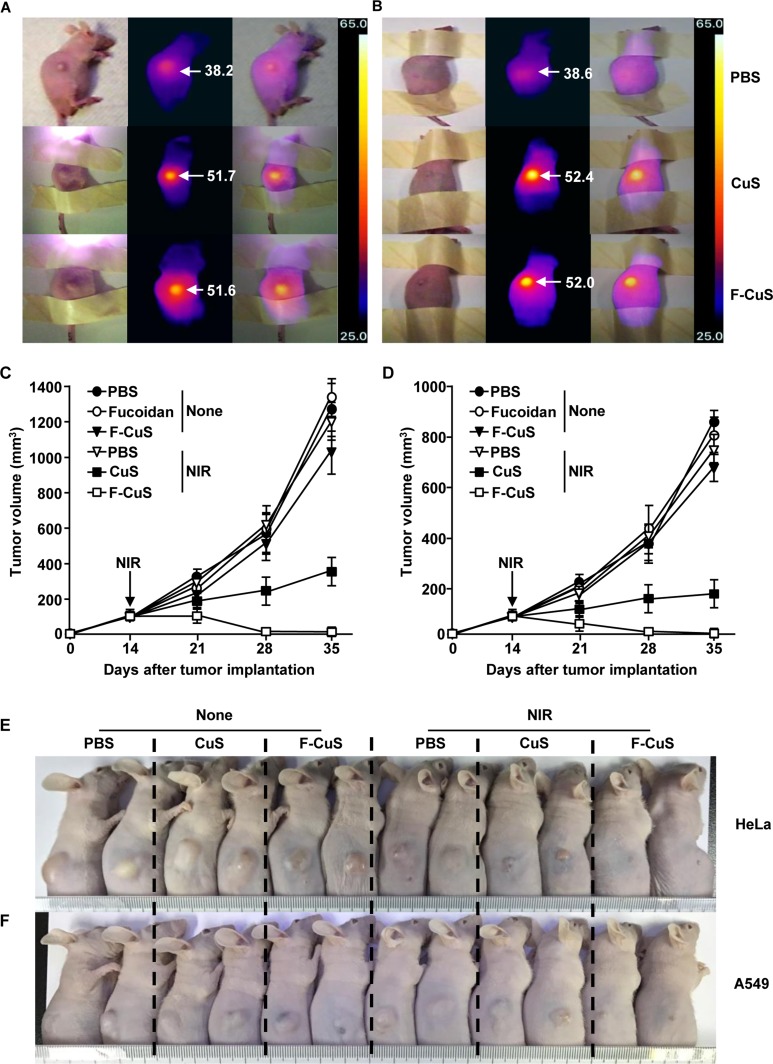
Figure 4. Chemo–photothermal therapy by F-CuS.
Nude mice were injected s.c. with 5 × 10⁶ HeLa cells and 5 × 10⁶ A549 cells. Once tumors were measured to be ~5.0 mm (after 14 d), the mice were treated i.t. with 4 μg/kg fucoidan, 10 mg/kg CuS, or 2.5 mg/kg F-CuS. Two hours after treatment, the mice were irradiated for 5 min with an 808 nm laser at 2 W/cm2. (A and B) Thermal image of HeLa tumor mice (A) and A549 tumor mice (B) are shown after NIR irradiation. (C and D) Tumor volumes of mice injected with HeLa (C) and A549 (D) were measured. (E and F) Tumor masses in the mice are shown after the mice were sacrificed on day 35 of HeLa (E) and A549 (F) cell xenograft. All data are representative of, or the average of, analyses of six independent samples (i.e., two samples per experiment, three independent experiments).

