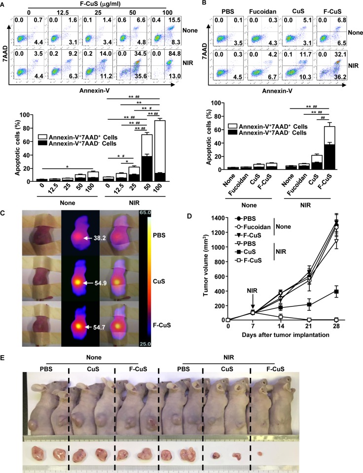
Figure 5. F-CuS-mediated chemo–photothermal therapy against multi-drug-resistant K562 cells.
(A) K562 cells (2 × 10⁵) were treated with an indicated dose of F-CuS for 2 h; cells were irradiated for 5 min with an 808 nm laser at 2.5 W/cm2. Apoptosis of K562 cells was analyzed by annexin-V & 7AAD staining (upper panel). (B) K562 cells were treated with 75 ng/mL fucoidan, 200 μg/mL CuS, or 50 μg/mL F-CuS for 2 h, and then the cells were irradiated for 5 min with an 808 nm laser at 2.5 W/cm2. Apoptotic K562 cells are shown (upper panel). In (A) and (B), the mean percentages of early-apoptotic cells (annexin-V+7AAD– cells) and late-apoptotic/necrotic cells (annexin-V+7AAD+ cells) are shown (right panel). #p < 0.05, ##p < 0.01 for early-apoptotic cells; *p < 0.05, **p < 0.01 for late-apoptotic/necrotic cells (lower panel). (C to E) Nude mice were injected s.c. with 5 × 10⁶ K562 cells. Once tumors measured to be ~5.0 mm (after 14 d), the mice were treated i.t. with 4 μg/kg fucoidan, 10 mg/kg CuS, or 2.5 mg/kg F-CuS. Two hours after treatment, the mice were irradiated for 5 min with an 808 nm laser at 2.5 W/cm2. (C) Thermal images of mice are shown after NIR irradiation. (D) Tumor volumes are shown. (E) Tumor masses in the mice are shown on day 28 of NIR irradiation. Data are representative of analyses of four independent samples (i.e., two mice per experiment, two independent experiments).

