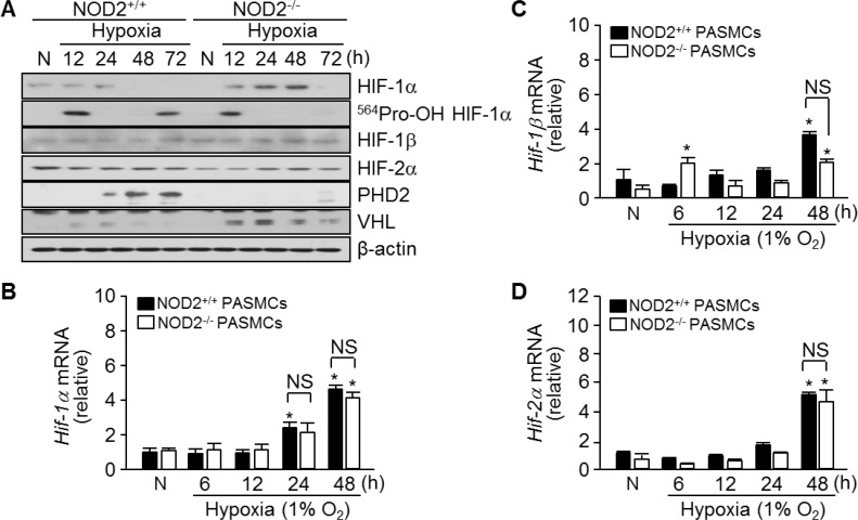
Figure 6. Absence of NOD2 enhances the stability of HIF-1α protein in PASMCs exposed to hypoxic conditions.
(A) Total protein was extracted from NOD2+/+ and NOD2−/− PASMCs exposed to normoxic (N) or hypoxic conditions for the indicated lengths of time. The protein levels of HIF-1α, hydroxylated HIF-1α (Pro564), HIF-1β, HIF-2α, PHD2 and VHL were then assessed by Western blot; β-actin was used as a loading control. Experiments were performed at least three independent times. (B–D), Total RNA was extracted from NOD2+/+ and NOD2−/− PASMCs exposed to normoxic (N) or hypoxic conditions for the indicated lengths of time. The mRNA levels of Hif-1α (B), Hif-1β (C) and Hif-2α (D) were then analyzed by quantitative real-time RT-PCR; mouse β-actin was used as a control for normalization. *P < 0.05, upregulation after hypoxia vs. after normoxia (N). NS, not significant. Values are presented as means ± SDs, n = 3.

