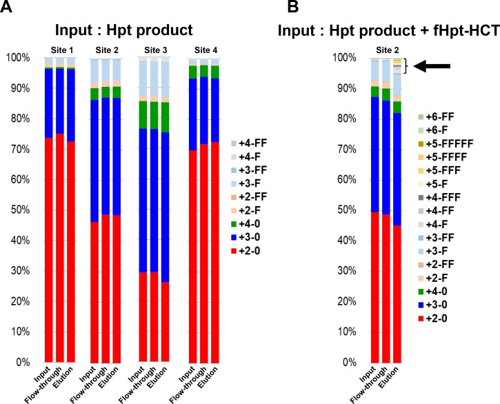
Figure 3. Affinity chromatography with the 10-7G mAb followed by site-specific analyses of N-glycans on haptoglobin by mass spectrometry.
The ratio of various N-glycans at each site on haptoglobin was calculated from the results of mass spectrometry for three fractions (input, flow-through, elution) under affinity chromatography with the 10-7G mAb. Hpt products (A) and a sample consisting of a mixture of Hpt products + 1% fHpt-HCT purified from Hpt-transfected HCT116 cells (B) were used in chromatography. The arrow indicates highly branched N-glycans on Fuc-Hpt and/or proHpt derived from HCT116 cells. Site 1: Asn 184; Site 2: Asn 207; Site 3: Asn 211; Site 4: Asn 241. For the abbreviations of N-glycan structures on the glycopeptides, the simplified notation was used. For example, the peptide containing the tri-antennary N-glycan with one Fuc residue is represented as “3-F.” The first numeral indicates the branch number (tri-antennary in this case) or LAcNAc number (3 LAcNAc in this case), and “F” indicates one Fuc residue. “0” denotes the absence of Fuc.

