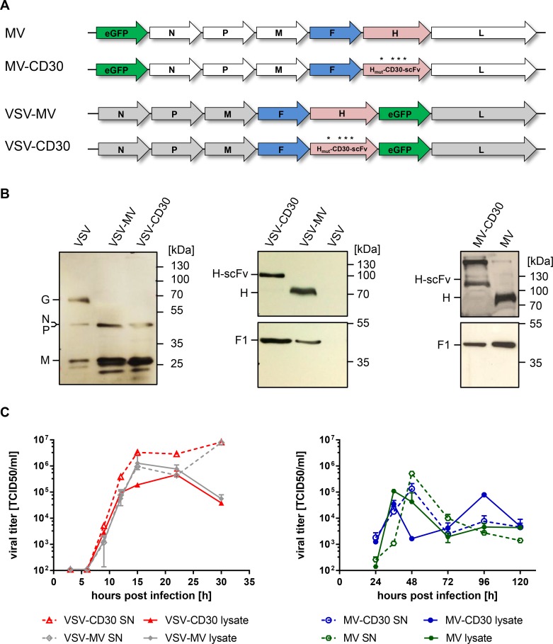
Figure 1. Generation of MV-CD30 and VSV-CD30.
(A) Schematic genome organization of the applied oncolytic viruses. Asterisks indicate the mutated residues in H protein to achieve blinding for the natural MV receptors CD46 and SLAM. The coding sequence for the CD30-specific scFv together with a C-terminal hexa-His-tag (H6) is fused to Hmut. In VSV-CD30 the glycoprotein (G) reading frame was replaced with those of MV-F and Hmut-CD30scFv (B) Immunoblot of virus stocks using rabbit-α-VSV serum (left panel) or antibodies recognizing the cytoplasmic tail of MV-H (center and right panel top row) or MV-F (center and right panel bottom row). (C) Multi-step growth curves of CD30-targeted viruses and untargeted parental viruses on Vero-αHis cells of cell-associated (lysate) and supernatant (SN) virus after infection at a multiplicity of infection (MOI) of 0.3, respectively. Titers were determined as 50% tissue culture infective dose (TCID50). n = 2, error bars: mean ± SD.

