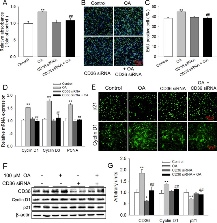
Figure 2. Knockdown of CD36 eliminated the enhancement of HC11 proliferation induced by OA.
(A, B) Effects of 100 μM OA and/or CD36 siRNA on HC11 proliferation was determined using MTT analysis (A) and EdU incorporation assay (B). The nuclei were stained with Hoechst and the scale bar = 200 μm. (C) Analysis of EdU positive cell percentage in panel B. (D) The relative mRNA expression level of Cyclin D1, Cyclin D3, and PCNA in response to 100 μM OA and/or CD36 siRNA. (E) The representative immunofluorescence staining of Cyclin D1 and p21 in the presence of 100 μM OA and/or CD36 siRNA. Scale bar = 100 μm. (F) Western blot analysis of CD36, Cyclin D1 and p21 in HC11 after a 4-day culture. β-actin was used as the loading control. (G) Mean ± SEM of immunoblotting bands of CD36, Cyclin D1, and p21. The intensities of the bands were expressed as the arbitrary units. **P < 0.01 versus the Control group, ##P < 0.01 versus the 100 μM OA group.

