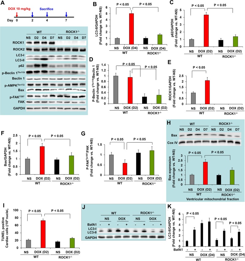
Figure 5. ROCK1 deletion inhibited the early onset of doxorubicin-induced autophagy dysregulation and apoptosis.
(A). Schematic of single DOX administration protocol (top). Mice 8 to 9 weeks old received one injection of NS or DOX (10 mg/kg). Mice were sacrificed on day 2, 4 and 7 after the injection. Representative images (bottom) of Western blot analysis of ROCK1, ROCK2, LC3, p62, Beclin 1, p-Beclin 1-Thr119, p-AMPK-Thr172, Bax, FAK and p-FAK-Tyr397 in ventricular homogenates of WT and ROCK1 deficient hearts on day 2, 4 and 7 after a single DOX injection. (B-G). Quantitative analysis of immunoreactive bands of LC3-II on day 4 (B), p62 on day 2 (C), p-Beclin1-Thr119/Beclin 1 on day 2 (D), ROCK1 on day 2 (E), Bax on day 2 (F), p-FAK-Tyr397/FAK on day 2 (G) after single DOX injection. N = 4-6 in each group. (H). Representative images (top) of Western blot analysis of Bax and Cox IV in the mitochondrial fraction of ventricular homogenates from WT and ROCK1 deficient hearts. Quantitative analysis (bottom) of immunoreactive bands of Bax on day 2 after single DOX injection (N = 4-6 in each group) expressed as fold change relative to NS-treated WT group. (I). Quantification of total TUNEL positive nuclei per 105 total nuclei in ventricular myocardium from WT and ROCK1 deficient hearts on day 2 after single DOX injection. N = 4-6 in each group. (J-K). Mice 8 to 9 weeks old received one injection of saline or DOX (10 mg/kg). On day 4 after the injection, mice received one injection of bafilomycin A1 at 1.5 mg/kg 2 hours before they were sacrificed. Representative images (J) and quantitative analysis (K) of Western blot analysis of LC3-II in ventricular homogenates of WT and ROCK1 deficient hearts. N = 4-6 in each group expressed as fold change relative to NS-treated WT group.

