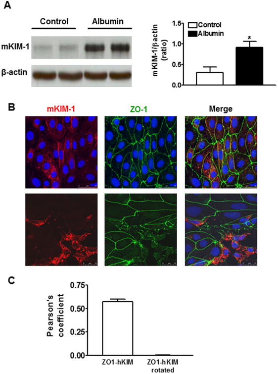
Fig. 1.

KIM-1 protein expression in primary cultures of mouse tubular epithelial cells (TECs) following albumin stimulation. A: KIM-1 protein level was significantly increased when the primary mouse renal TECs were incubated with bovine serum albumin (10 mg/ml) for 72 h. An n of 3–4 epithelial cultures were treated for each condition; *P < 0.05 versus untreated control group. B: Double immunostaining for mKIM-1 and Zonula Occludens (ZO-1) show that mKIM-1 red staining was present at the cell-cell border (green ZO-1) and in small intracellular vesicles in untreated primary TECs (top). Following albumin stimulation for 72 h, mKIM-1 was concentrated in large perinuclear vesicles in association with a disruption of cell–cell contacts (bottom). Images are Z-stack projections. C: Quantification of co-localization of images as in panel B top using Pearson's correlation coefficient (PCC). The negative control was provided by quantifying PCC for the same images, but after rotation of one by 90°. The results represent more than 20 cells from at least n = 2 independent experiments.
