
Fig. 5.
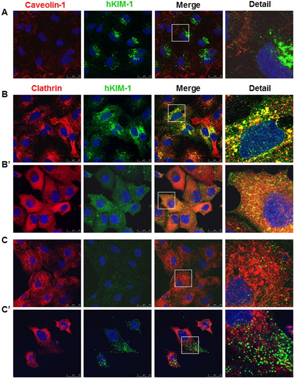
Colocalization of KIM-1 with clathrin in renal tubular epithelial cells. A: There was no significant colocalization between KIM-1 and caveolin-1 in HK-2 cells. B: A colocalization of KIM-1 and clathrin was evident in the perinuclear area of HK-2 cells. Removal of AP2 from the plasma membrane by chlorpromazine markedly increased the lateral diffusion of clathrin and KIM-1 (B′) compared to their predominant perinuclear localization in the absence of chlorpromazine (B). C–C′: In non-permeabilized HK-2 cells, chlorpromazine administration increased the number and size of KIM-1 puncta on the cell surface (C′) compared to untreated cells (C). Staining was repeated at least three times with similar results. Images are Z-stack projections.