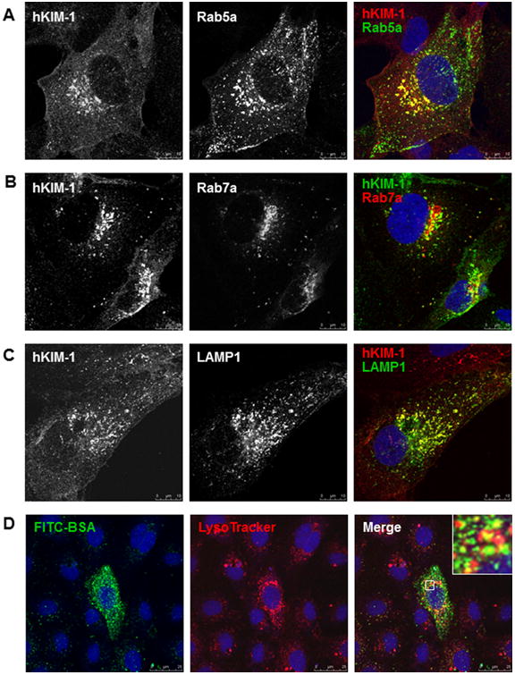
Fig. 7.

Endocytic trafficking of endogenous KIM-1 in HK-2 cells. A–C: HK-2 cells were transfected with a plasmid encoding GFP-Rab5a, RFP-Rab7a, or GFP-LAMP1 for 16 h and then immunostained with a goat anti-hKIM-1 antibody. Representative confocal images show colocalization of KIM-1 with early endosomes (A, Rab5a), late endosomes (B, Rab7a), and lysosomes (C, LAMP1). D: hKIM-1-transfected NRK-52E cells were incubated with FITC-BSA (green) and LysoTracker (red) for 30 min at 37°C. A distribution of BSA in acidic lysosomal compartments was evident in the merged image (yellow). Staining was repeated three times with similar results. Images are Z-stack projections.
