
Fig. 8.
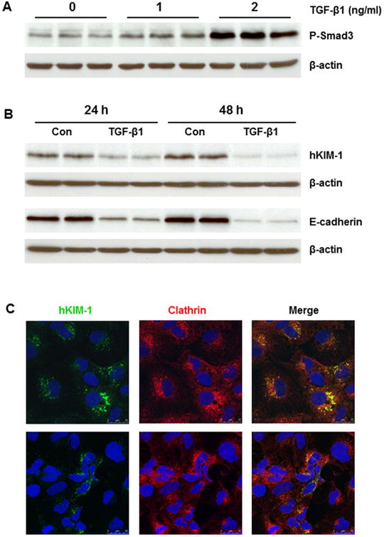
Effect of TGF-β1 on KIM-1 protein in HK-2 cells. A: TGF-β1 (1 and 2 ng/ml) led to a dose-dependent increase of Smad3 phosphorylation in HK-2 cells after 24 h. B: TGF-β1 (2 ng/ml) significantly reduced hKIM-1 and E-cadherin protein levels after 24 and 48 h. C: Representative confocal images show that hKIM-1 staining was reduced in HK-2 cells treated with TGF-β1 (2 ng/ml) for 24 h (bottom) compared to untreated cells (top).