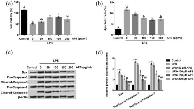
Figure 2.
Effects of APS on LPS-induced inflammation injury in H9c2 cells. H9c2 cells were treated with LPS or co-treated with APS and LPS for 24 h. (a) Cell viability was evaluated by CCK-8 assay. (b) Cell apoptosis of H9c2 cells was detected by flow cytometry. (c and d) The apoptosis-related protein levels were examined by western blot. Different letters above the bars (a, b, c, d) indicate that the means of different groups were significantly different (P < 0.05) by ANOVA. Each experiment was repeated at least three times.
APS: Astragalus polysaccharide; LPS: lipopolysaccharide; CCK-8:Cell Counting Kit-8; ANOVA: one-way analysis of variance.
***P < 0.001 vs control group; #P < 0.05, ##P < 0.01, ###P < 0.001 vs LPS group.

