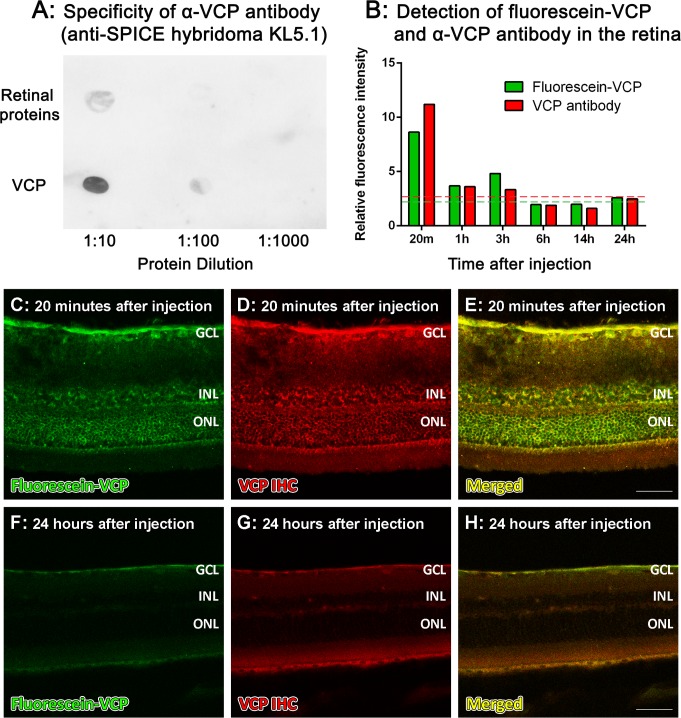
Fig 4. Localisation of VCP in the retina following an intravitreal injection.
A: Dot blot analysis showed immunoreactivity of the α-VCP antibody (anti-SPICE hybridoma KL5.1) for VCP, compared to mixed retinal proteins (control). B-H: 10μg of fluorescein-VCP was intravitreally injected into the retina, and retinal fluorescence intensity (B) was determined from 20 minutes to 24 hours after injection. The red dashed line represents background red fluorescence levels in negative control and non-injected control sections, and the green dashed line represents background green fluorescence levels of non-injected control sections. Fluorescein-VCP was detected in all layers of the retina, and was evenly distributed from the central retina to the periphery at 20 minutes (C). However, at 1–3 hours after injection, the fluorescein-VCP was cleared from the retina, which remained at background levels of fluorescence up to 24 hours (F). Double labelling with the α-VCP antibody was used to verify localisation of VCP in the retina (B, D, E, G, H). GCL, ganglion cell layer; INL, inner nuclear layer; ONL, outer nuclear layer; h, hours; m, minutes. Scale bars indicate 50μm.

