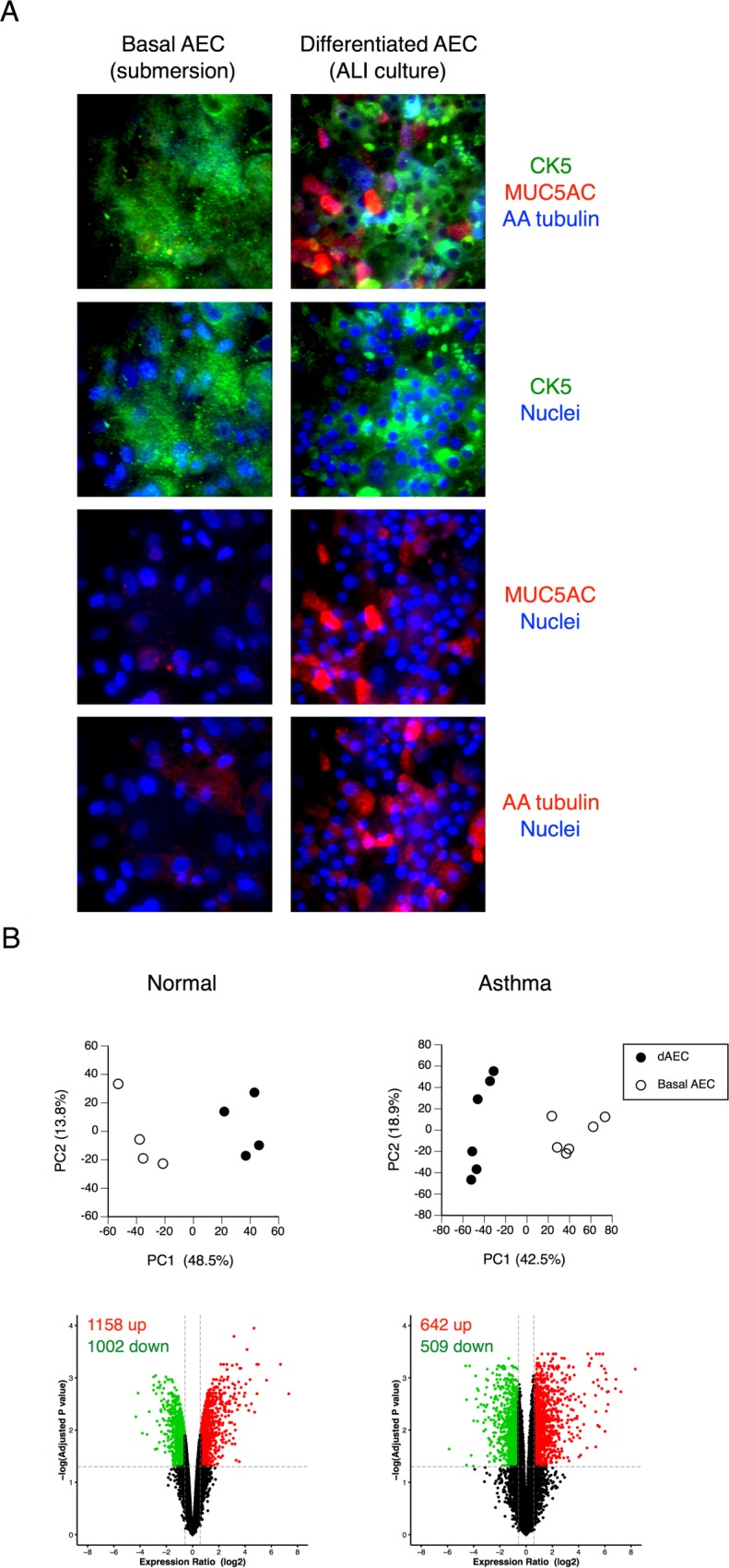
Fig 1. Demonstration of airway epithelial cell differentiation.

A. Confocal immunofluorescence of basal and differentiated (dAEC) to demonstrate markers of differentiation in the latter. The presence of MUC5AC indicated differentiation into goblet cells, and the presence of alpha-acetylated tubulin indicated differentiation into ciliated cells. The presence of CK5 indicated the presence of basal cells in both culture conditions. No significant differentiation into goblet or ciliated cells types is seen in cells grown in submersion culture. B. Principal component analysis and volcano plots in AEC from normal or asthmatic donor lungs to demonstrate differential gene expression in the resting (without mechanical injury) state (N = 4 in each group) between dAEC and basal AEC, using all expressed gene probe sets (n = 35,530 for normal cells and n = 41,733 for asthmatic cells) as an input dataset. For each, the number of up- and down-regulated probes (≥1.5 fold change, vertical dashed lines, adjusted P < 0.05, horizontal dashed line) is provided.
