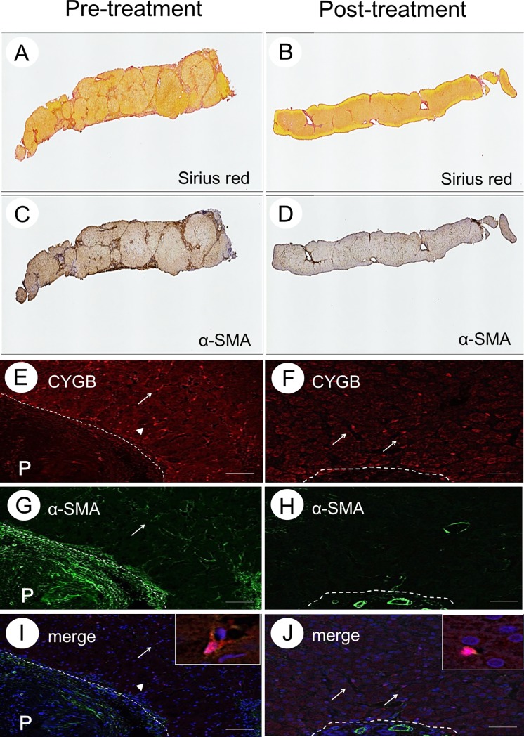
Fig 2. Collagen deposition and the presence of α-SMA-positive cells in liver tissue without HCC.
Sirius red staining (A and B), α-SMA immunohistochemistry (C and D), CYGB immunofluorescence staining (E and F), α-SMA immunofluorescence staining (G and H), merged image of CYGB and α-SMA (I and J) of the liver specimen obtained before IFN treatment and after SVR for Case 1. Insets show enlarged views of CYGB/α-SMA-double-positive cells. Bar, 50 μm; P, portal vein.

