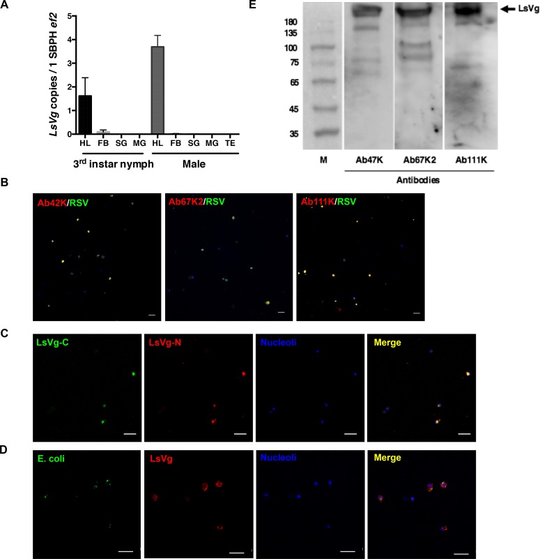
Fig 5. LsVg expression in SBPH nymphs and males.
A. LsVg mRNA distribution in different tissues of SBPH nymphs and males was revealed by qPCR. The mean and SD were calculated from three independent experiments, with four mRNA samples per experiment. Ef2, L. striatellus elongation factor 2 gene; HL, hemolymph; FB, fat body; SG, salivary glands; MG, midgut. TE, testis. B. Confocal microscopic image showing the existence of LsVg and its co-localization with RSV in hemocytes. LsVg was probed with LsVn-subunit specific antibodies Ab42K, Ab67K2 and Ab111K and stained with Alexa Fluor 568 (shown in red). RSV was stained with Alexa Fluor 488 (shown in green). Nucleoli were stained with TO-PRO-3 (shown in blue). C. Confocal microscopic image showing co-localization of the N-terminal small (Small) and C-terminal large (Large) subunits of LsVg. The large subunit was probed with antibody Ab111Km and stained with Alexa Fluor 488 (shown in green). The small subunit was probed with antibody Ab42K and stained with Alexa Fluor 568 (shown in red). D. Confocal microscopic image showing localization of LsVg and phagocytosed E. coli containing the gfp gene conferring green fluorescence. LsVg was probed with Ab42K and stained with Alexa Fluor 568 (shown in red). Images were examined using a Leica TCS SP8 confocal microscope. The scale bar represents 20 μm. E. LsVg is not cleaved in male L. striatellus. Extracted hemolymph proteins were fractionated by SDS-PAGE (10%) and probed with the subunit-specific antibodies Ab47Km, Ab67K2 and Ab111K. M, the molecular weight marker (kDa). Arrows on the right, identified LsVg proteins.

