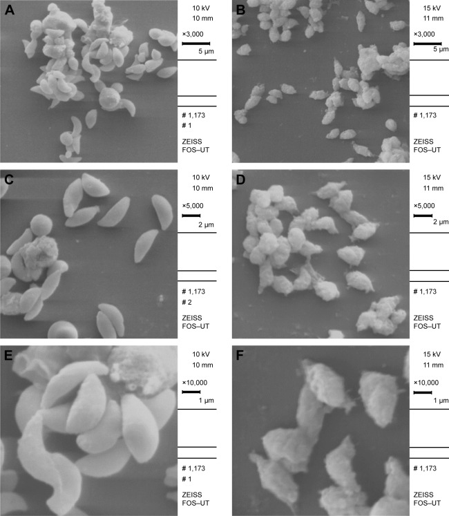
Figure 3.
(A, C and E) SEM micrographs of T. gondii tachyzoites in control group showing crescent-shaped parasites with obvious conoid (3,000×, 5,000× and 1,000× magnification, respectively). (B, D and F) SEM micrographs of T. gondii tachyzoites in group treated with 2,000 ppm of LMW CS NPs showing multiple deep ridges, irregular papules and large projections on the surface (3,000×, 5,000× and 1,000× magnification, respectively).
Abbreviations: SEM, scanning electron microscopy; LMW CS NPs, low molecular weight chitosan nanoparticles; T. gondii, Toxoplasma gondii.

