Figure 2.

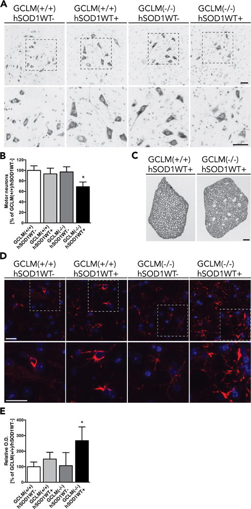
Motor neuron loss and astrogliosis in GCLM(−/−)/hSOD1WT+. A) Representative images from the lumbar ventral horn of 60 weeks old GCLM(−/−)/hSOD1WT+ mice and age-matched controls stained with cresyl violet. Higher magnification images of the indicated areas are shown in the lower panel. Scale bars: 50 μm. B) Number of large neurons in the ventral horn of the lumbar spinal cord from 60 weeks old mice of the indicated genotype. Data are presented as percentage of GCLM(+/+)/hSOD1WT− animals (mean±SD; n=4–5. * p<0.05). C) Luxol fast blue staining in cross sections of lumbar spinal cord roots from 60 weeks old GCLM(−/−)/hSOD1WT+ and age-matched GCLM(+/+)/hSOD1WT+ mice. Scale bar: 50 μm. D) Immunofluorescence against GFAP (red) in the anterior horn of lumbar spinal cord sections from 60 weeks old mice of the indicated transgenic genotypes. Nuclei were counterstained with DAPI. Higher magnification images of the indicated areas are shown in the lower panel. Scale bars: 20 μm. E) Relative optical density (O. D.) quantification of GFAP immunoreactivity in the grey matter of the anterior horn of lumbar spinal cord sections from the indicated genotypes. Data are presented as percentage of GCLM(+/+)/hSOD1WT− sections (mean±SD; * p<0.05)
