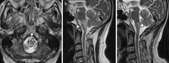
Figure 1.
Magnetic resonance imaging (MRI). (a) (b) Edematous changes of the lower brainstem are shown on axial and sagittal T2-weighted images. Abnormal flow voids in the subdural space are also seen (arrowheads). (c) A follow-up MRI study shows the resolution of brainstem congestion, while an infarction was observed to have developed at the dorsal medulla oblongata (arrowhead).

