Figure 2.

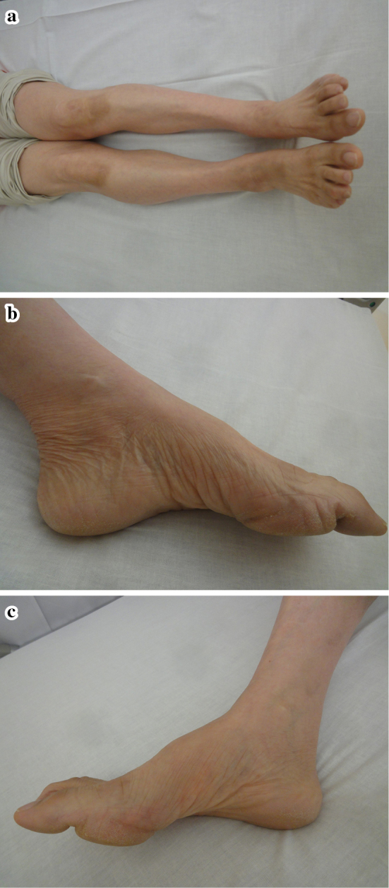
a-c: The patient showed distal dominant atrophy of both legs, including pes cavus. These features are often seen in Charcot-Marie-Tooth disease.

a-c: The patient showed distal dominant atrophy of both legs, including pes cavus. These features are often seen in Charcot-Marie-Tooth disease.