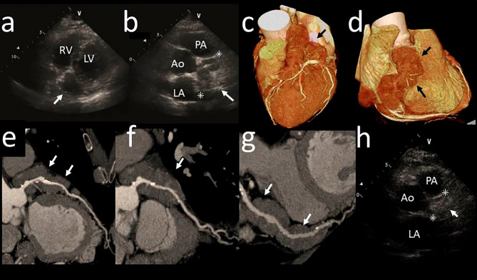
Figure 1.
Transthoracic echocardiography and computed tomography (CT) of the masses surrounding the coronary arteries. Transthoracic echocardiography depicted hypoechoic areas surrounding the left (a; parasternal short-axis view) and right (b; apex long-axis view) coronary arteries before treatment. Coronary CT angiography depicted diffuse masses surrounding the left (c) and right coronary arteries (d). No stenotic lesions or aneurysms were found in the left anterior descending (e), left circumflex coronary (f), or right (g) coronary arteries. Echocardiography showed that the hypoechoic areas were reduced in the right coronary artery (h) after treatment. Arrows denote the masses surrounding coronary arteries. Asterisks denote the maximum diameter. Ao: aorta, PA: pulmonary artery, LA: left atrium, LV: left ventricle, RA: right atrium

