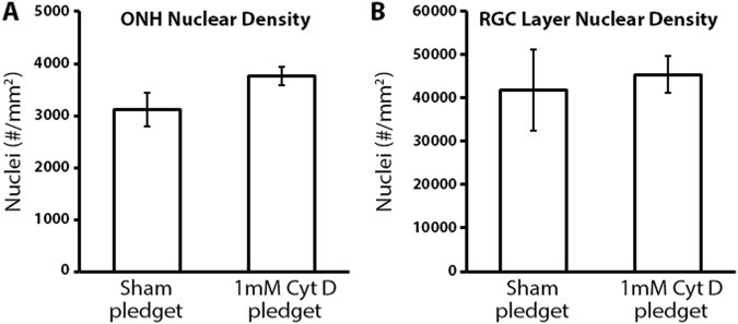
Figure 2.
The effect of cytochalasin D delivery on optic nerve head and retinal ganglion cell layer nuclear counts. (A) Nuclear density within the anterior optic nerve head (ONH; 5 µm sections; 0–100 µm posterior to Bruch’s membrane) after sham (n = 4) and cytochalasin D (Cyt D; n = 7) delivery to the optic nerve (no statistically significant difference was noted between groups). (B) Nuclear density within the retinal ganglion cell (RGC) layer of the superior peripapillary retina (5 µm sections; 0–250 µm from the superior optic nerve head) after sham (n = 4) and Cyt D (n = 6) delivery to the optic nerve (no statistically significant difference was noted between groups). Error bars indicate standard error of the mean.

