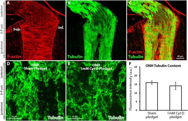
Figure 3.
The effect of cytochalasin D delivery on optic nerve head axonal microtubule levels. (A–C) Normal optic nerve head (ONH) labeled with fluorescent-tagged phalloidin (F-actin maker) and axon-specific anti-βIII tubulin antibodies. (D,E) ONH tubulin levels after sham (pledget soaked in vehicle only) delivery and 1 mM Cyt D delivery to the junction of the superior optic nerve and globe. (F) ONH tubulin content assessed by fluorescence intensity measurement of anti-βIII tubulin antibody labeling after sham (n = 4) and 1 mM Cyt D delivery (n = 6) to the optic nerve (no statistically significant difference was noted between groups). Error bars indicate standard error of the mean. a.u. = arbitrary units; Cyt D = cytochalasin D; F-actin = filamentous actin; Inf. = inferior; ONH = optic nerve head; Sup. = superior.

