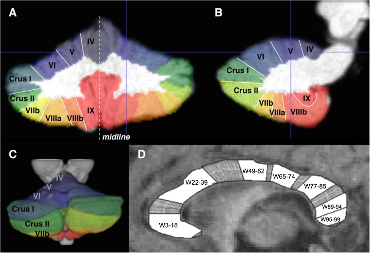
Fig. 1.
Parcellation of cerebellum and corpus callosum. a–c Automatic segmentation results using the SUIT toolbox in SPM8. Different colours mask individual lobule volumes in anatomical MRI space in a coronal, b sagittal and c 3D-reconstructed views. Crosshairs show right lobule V. d Midsagittal view of the corpus callosum (anterior = left of image) with regional clusters depicted in relation to the 99 percentile widths

