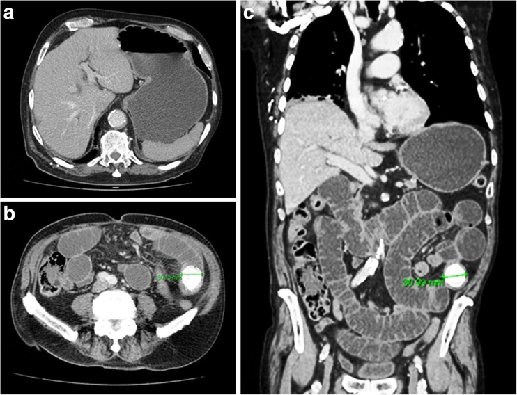
Fig. 2.
Contrast CT abdomen/pelvis images at time of admission. a CT slice demonstrating a dilated, fluid-filled stomach. Nil significant pneumobilia noted. b, c CT slices demonstrating an obstructing, calcified mid-jejunal intraluminal stone measuring 3 cm in diameter. Evidence of small bowel obstruction noted with dilated jejunal loops above the obstruction. Colon is of normal caliber

