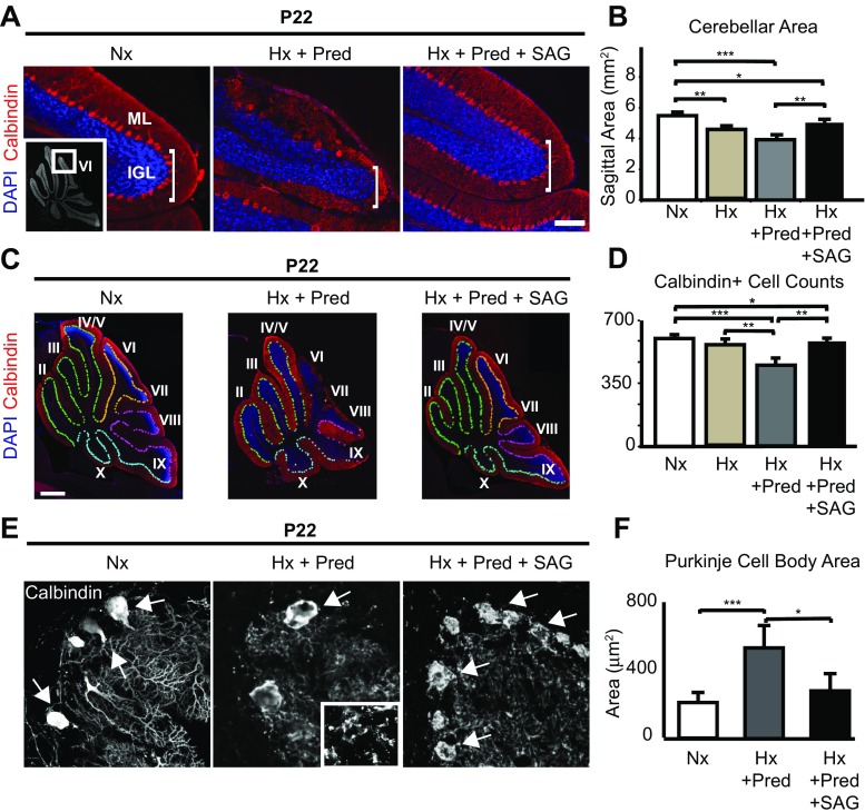
Fig. 3.
Chronic hypoxia plus prednisolone result in increased cerebellar hypoplasia and Purkinje cell loss, and SAG can partially rescue both phenotypes. a Representative images of lobule 6 in the cerebellar vermis showing the molecular layer (ML) and internal granule layer (IGL) from normoxic (Nx), hypoxic plus prednisolone (Hx + Pred), or hypoxic plus prednisolone and SAG (Hx + Pred + SAG) brain samples. Purkinje cells stained in red (Calbindin), and nuclei counterstained in blue (DAPI). Note size difference of IGL among the samples. Insert, location of lobule 6 as shown in whole cerebellum at lower magnification. Scale bar, 50 μm. b Quantification of cerebellar vermis area in Nx, Hx + Pred, and Hx + Pred + SAG conditions. Nx = 5.49 ± 0.104 mm2 (n = 3), Hx = 4.56 ± 0.120 mm2 (n = 3), Hx + Pred = 3.93 ± 0.324 mm2 (n = 5), Hx + Pred + SAG = 5.03 ± 0.062 mm2 (n = 5). c Representative images of whole cerebella showing extent of Purkinje cell damage in Nx, Hx + Pred, and Hx + Pred + SAG brains at P22. Purkinje cells stained with Calbindin (red), nuclei counterstained with DAPI (blue). Pseudocolors show demarcation of Purkinje cells from anterior (light green) through medial (orange/pink) and posterior (light blue) regions. Scale bar, 1 mm. d Quantification of Calbindin positive cells in Nx, Hx, Hx + Pred, or Hx + Pred + SAG. Nx = 604 ± 13 cells (n = 3), Hx = 562 ± 10 cells (n = 5), Hx + Pred = 452 ± 27 cells (n = 6), Hx + Pred + SAG = 535 ± 7 cells (n = 5). For quantification, mean + SEM. e Representative images of high-power Calbindin-positive Purkinje cells in Nx, Hx + Pred, or Hx + Pred + SAG brains at P22 showing extent of cell and dendritic damage. Arrows indicate Purkinje cell soma. Insert, higher power magnification of molecular layer showing disruption of dendrites and absence of Caspase 3. f Quantification of Purkinje cell body area. Nx = 216 ± 27.4 μm2, Hx + Pred = 551 ± 60.9 μm2, Hx + Pred + SAG = 290 ± 46.9 μm2. For each condition, n = 3 brains, average of 25 cells. *p < 0.05, **p < 0.01, ***p < 0.001, ANOVA with Tukey’s post-hoc correction

