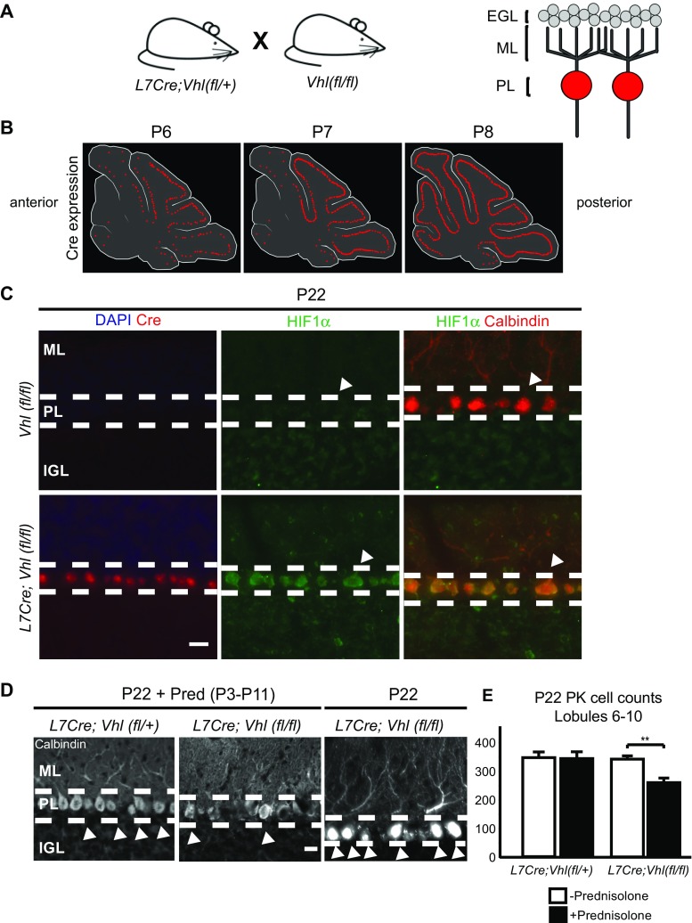
Fig. 5.
The HIF pathway plays a role in PK cell injury from prednisolone administration in L7Cre;Vhl(fl/fl) mice. a Right, schematic diagram showing transgenic mouse breeding to target Purkinje cells in L7Cre;Vhl(fl/fl) animals. Left, schematic of cerebellar circuit highlighting Purkinje-specific Cre recombination (red). EGL external granule layer, ML molecular layer, PL Purkinje cell layer. b Diagram highlighting timeline of Cre expression in P6 to P8 mouse pups. Note Cre expression turns on in posterior regions, specifically in lobules 6–9 of the cerebellar vermis, before anterior regions. c Representative images showing Cre (red, left column) and HIF1α (green) expression in the PL at P22 only in L7Cre;Vhl(fl/fl) animals. Note HIF1α colocalization with Calbindin (red, right column, denoted by arrowheads) expression in the PL. ML molecular layer, PL Purkinje cell layer, IGL internal granule layer. Scale bar, 20 μm. d Representative images showing loss of Calbindin + cells (arrowheads) in P22 L7Cre;Vhl(fl/fl) animals given daily Pred injections from P3 to P11. Dashed lines denote layer borders similar to (c). Scale bar, 10 μm. e Quantification of Calbindin + PK cells in posterior lobules. L7Cre;Vhl(fl/+) = 343.3 ± 19.6 cells (n = 3), L7Cre;Vhl(fl/fl) = 338.5 ± 23.5 cells (n = 3), L7Cre;Vhl(fl/+) + Pred = 339.2 ± 9.43 cells (n = 3), L7Cre;Vhl(fl/fl) + Pred = 255.8 ± 16.1 cells (n = 4). **p < 0.01, Student’s t test. For quantification, n ≥ 3 experiments per condition

