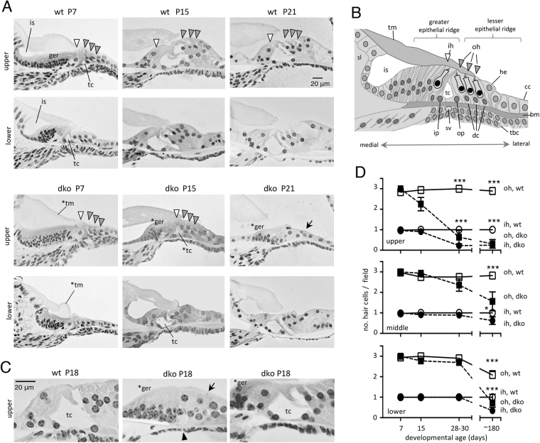
Figure 4.
Developmental and degenerative cochlear abnormalities in dko mice. (A) At P7, the cochlea is being remodeled: in wt, the greater epithelial ridge (ger) is partly regressed and the inner sulcus (is) and tunnel of Corti (tc) are partly open. Lower regions are more advanced than upper regions because of the basal-apical progression of development. At P15, remodeling nears completion with upright, elongated hair cells (arrowheads) and open tc. The dko at P7 has hair cells and support cells but has almost no opening of the inner sulcus or tc and a malformed tectorial membrane (*tm). The dko is still retarded at P15 with limited regression of the ger (*ger) and poor opening of the tc (*tc), especially in upper regions. At P21, degeneration of the sensory epithelium (arrow) is also evident in upper regions. (B) Diagram of cell types of the immature cochlea (at ~P7). Abbreviations: bm, basilar membrane; cc, Claudius cells; dc, Deiters cells; he, Hensen cells; ih, inner hair cell; ip, inner pillar cell; is, inner sulcus; oh, outer hair cell; op, outer pillar cell; sl, spiral limbus; sv, spiral vessel; tbc, tympanic border cell; tc, tunnel of Corti; tm, tectorial membrane. The spiral vessel below the bm is prominent at birth and regresses during postnatal development59. (C) Higher magnification showing phenotypes in dko mice at P18. A mild example (right) shows delay in regression of ger cells (*ger) but an open tc, resembling wt (left). A severe example (middle) has delayed regression of the ger, a degenerated sensory epithelium (arrow) and absent tc (arrowhead). (D) Counts of outer (oh) and inner (ih) hair cells indicate the progression of degeneration in dko mice from P7 to P180 (6 months), determined on histological sections (mean ± sem). For wt versus dko at indicated ages: *p = 0.0396, ***p < 0.001.

