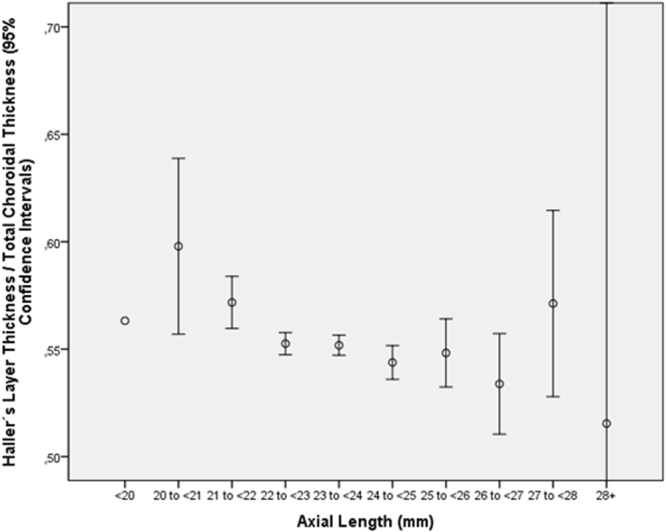
Figure 7.

Graph showing the distribution of the ratio of thickness of the subfoveal large choroidal vessel layer (Haller’s layer) to total subfoveal choroidal thickness, stratified by axial length, in the Beijing Eye Study 2011 in eyes without glaucoma, age-related macular degeneration, diabetic retinopathy, retinal vein occlusions, polypoidal choroidal vasculopathy or central serous choroidopathy and with an axial length less than 26.5 mm.
