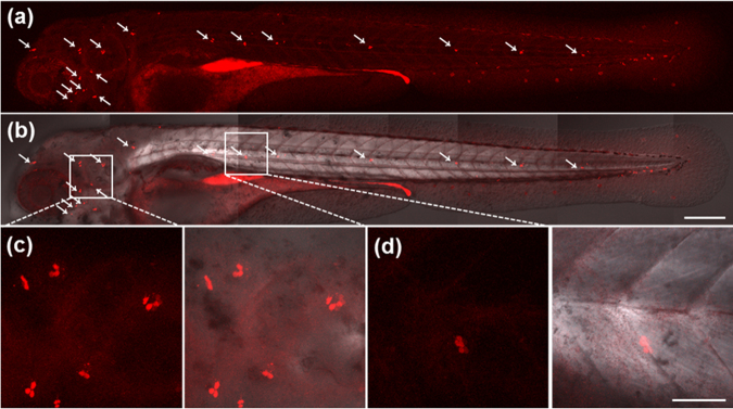
Figure 4.
Sensory hair cells in the lateral line of whole zebrafish larvae at 4 dpf. White arrows (a and b) show the lateral line hair cells in a zebrafish larva incubated with FPNPs (2 mg mL−1) from 2 to 4 dpf. (a) Fluorescence image, (b) Merged image with DIC. Enlarged images of (c) cranial neuromasts and (d) trunk neuromasts. The fluorescence was determined at 543 nm with excitation at 405 nm. Scale bars: 200 μm (a,b), 50 μm (c,d).

