Figure 4.

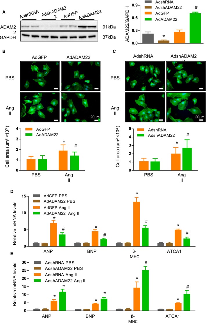
A disintegrin and metalloprotease‐22 (ADAM22) modulates angiotensin II (AngII)–induced cardiomyocyte hypertrophy in vitro. A, Western blot analysis of ADAM22 expression. B and C, Fluorescent analysis of neonatal rat cardiomyocytes (NRCMs) that had been infected with adenoviral vector encoding the green fluorescent protein gene (AdGFP) or overexpression ADAM22 (AdADAM22; B) or with nontargeting control (AdshRNA) or AdADAM22 (C), and treated with AngII (1 μmol/L) or PBS for 48 hours (blue, nuclear; green, α‐actinin; bar=20 μm). The cell surface areas in individual groups of cells were assessed (n>100 cells/group were examined). D and E, The relative levels of hypertrophic marker mRNA transcripts in NRCMs that had been infected with AdGFP or AdADAM22 (D) or with AdshRNA or knockdown ADAM22 expression (AdshADAM22; E) (n=7 samples per group). Data are representative images or present as the mean±SD of each group from at least 3 independent experiments. Statistical analysis was performed by 1‐way analysis of variance and post hoc tests. ANP indicates atrial natriuretic peptide; ATCA, acetyl‐Coenzyme A acetyltransferase 1; BNP, B‐type natriuretic peptide; and MHC, myosin heavy chain. *P<0.05 vs AdshRNA or AdGFP/PBS group; † P<0.05 vs AdshRNA or AdGFP/AngII group.
