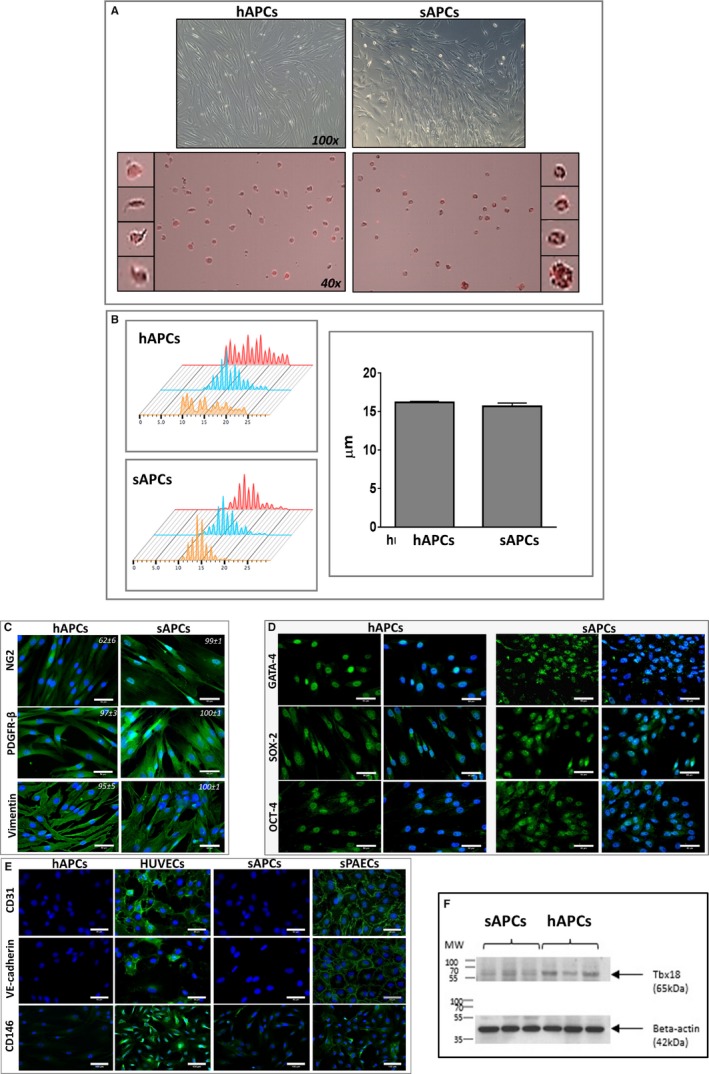
Figure 5.

Comparison of APCs isolated from human and swine saphenous veins. A, Upper panels: phase contrast microscopy images of human and swine cells displaying similar spindle‐shape features (magnification ×100). Lower panels: Cells shown in the contrast‐phase microscopy images are stained with swine and human PE CD105 antibodies (PE=red laser) for measurement of cell size by Tali Image‐based cytometer (magnification ×40). B, Cell size histograms calculated by Tali and Novocyte 3000 (Acea, Biosciences, Inc). The X axis in the left panels represents the cell size and the Y axis represents the number of cells counted (3 cell lines for each group). The bar graph shows the mean and SEM, which was similar between groups, though the range of cell size was wider for hAPCs. C, Representative immunofluorescence microscopy images show both hAPCs and sAPCs express the typical mesenchymal markers NG2, PDGFR‐β, and Vimentin. Values in each panel represent the mean±SEM of 4 biological replicates. D, Immunofluorescence microscopy images show the expression of cardiac transcriptional factor GATA‐4, and the stemness markers OCT‐4 and SOX‐2. Blue fluorescence of DAPI recognizes nuclei. Magnification ×200 and ×400 (50‐μm scale bar). E, Representative immunofluorescence microscopy images of hAPCs and sAPCs confirming these cells do not express endothelial antigens, at variance with HUVECs and PAECs (positive controls). F, Western blot image showing Tbx18 protein corresponding to the 65 kDa MW within human and swine APC lysate. APCs indicates adventitial pericytes; DAPI, 4′,6‐diamidino‐2‐phenylindole; GATA‐4, GATA binding protein 4; hAPCs, human adventitial pericytes; HUVECs, human umbilical vein endothelial cells; MW, molecular weight; sPAECs, swine pulmonary artery endothelial cells; PDGFR‐β, platelet‐derived growth factor receptor‐β; OCT‐4, octamer‐binding transcription factor 4; sAPCs, swine adventitial pericytes; SOX‐2, sex determining region Y‐box 2.
