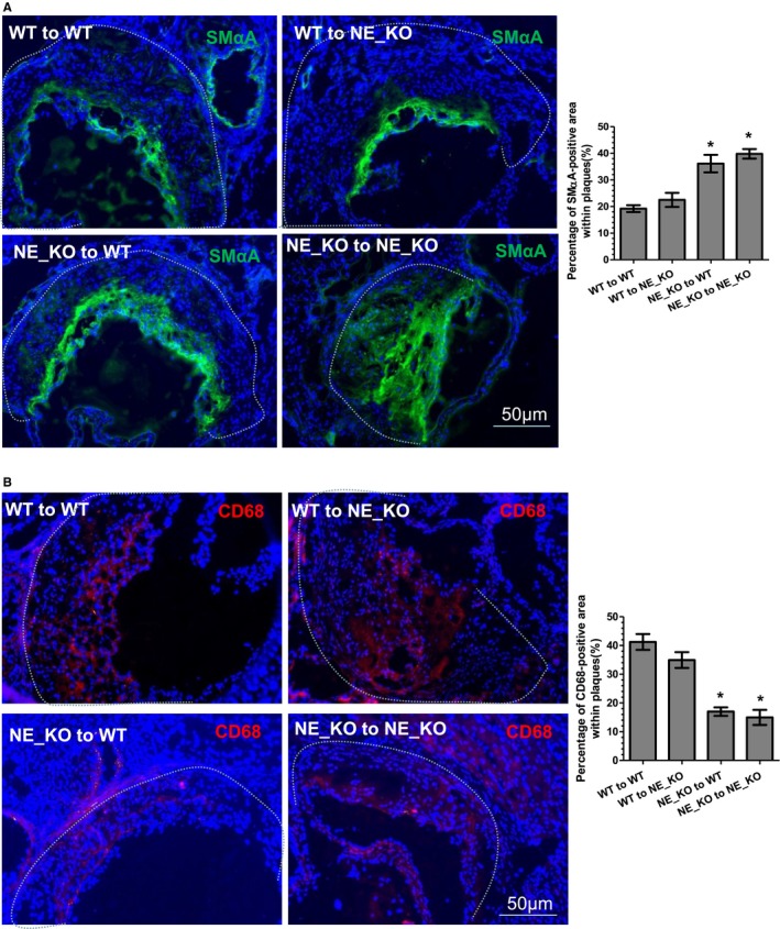
Figure 9.

Smooth muscle cells and macrophage content in atherosclerotic plaques of the mice with bone marrow transplantation. Aortic roots from 4 groups of transplanted mice were harvested and subjected to immunofluorescent staining analyses using antibodies against smooth muscle α‐actin (SMαA) (A) and CD68 (B), respectively. Respective representative images and quantitative data (bar graphs) from 10 mice (n=10 mice per group) are presented. Dot lines indicate the boundary between media layer and atherosclerotic lesion. *P<0.05 (vs wild‐type [WT] to WT mice). NE_KO indicates neutrophil elastase−/−/Apolipoprotein E−/− double knockout mice.
