Figure 5.

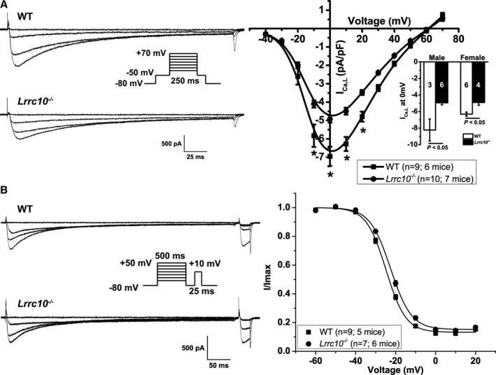
Lrrc10 −/− ventricular myocytes have reduced ICa,L and shifted steady‐state inactivation of ICa,L. A, ICa,L recordings from wild‐type (WT) or Lrrc10 −/− ventricular myocytes at −40, 0, 20, and +40 mV using the voltage protocol in the inset. Mean current–voltage (I‐V) relationship are plotted as mean±SEM *P<0.05. Inset shows mean peak ICa,L at 0 mV separated by sex with sample size shown in bars for 3 male WT mice, 6 female WT mice, 6 for Lrrc10 −/− male mice, and 4 Lrrc10 −/− female mice. B, Representative steady‐state inactivation traces at prepulses −60, −20, 0, and +20 mV. Mean V1/2 of inactivation was shifted to more depolarized potentials in Lrrc10 −/− myocytes compared with WT with data fitted by Boltzmann distributions.
