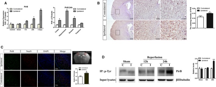
Figure 1.

A through D, Enhanced PirB expression and PirB signaling after transient middle cerebral artery occlusion (MCAO). A, Real‐time quantitative PCR analysis revealed highly increased PirB mRNA in the ischemic hemisphere compared with the undamaged contralateral side, sham‐operated hemisphere, and control group before MCAO both at 24 hours and at 4 days post‐MCAO (n=5). B, Representative images of immunohistochemical staining showed that PirB protein levels were markedly increased in the damaged hemisphere 24 hours post‐MCAO, compared with the undamaged contralateral side. Staining intensity of molecule was quantified by the simple PCI image analysis software (n=4). C, Majority of PirB immunostaining (green) colocalized with neuronal marker NeuN (red) in the cortical penumbra 24 hours after ischemia. Rectangle in T2‐weighted image indicates cortical area used for cell staining. Quantification of PirB staining in tMCAO brain slices was by ImageJ software (NIH, Bethesda, MD). Scale bar=100 μm (n=4). D, Protein extracts from hemispheres of post‐MCAO brains were subjected to immunoprecipitation for phosphotyrosine to assess levels of tyrosine phosphorylation of PirB. Expression of βIIItubulin was detected in input lysates. Phosphorylation of PirB was quantified as the ratio of band density to that of βIIItubulin. C indicates contralateral to ischemia; I, ipsilateral to ischemia; Sham, sham‐operated mice (n=4). Data are the mean±SEM. Statistical significance is indicated by: *P<0.05; **P<0.01. NeuN indicates neuronal nuclei; PirB, paired immunoglobulin‐like receptor B.
