Figure 2.

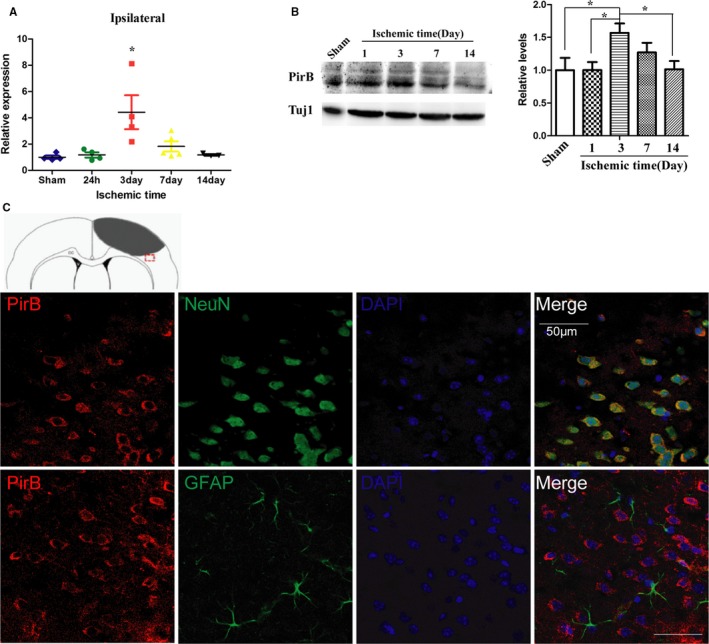
A through C, Expression and cellular location of PirB after cerebral cortex ischemia. Expression of PirB mRNA (A) and protein level (B) in the cortex with ischemia at different time points were detected by real‐time quantitative PCR and western blot analysis, respectively (n=4). Data are the mean±SEM. Statistical significance is indicated by *P<0.05, the expression level of ischemic cortex at different time points post‐injury compared with sham‐operated cortex. C, Representative images of PirB (red) and NeuN (green), or PirB (red) and GFAP (astrocytes, green) double immunofluorescene staining in the ischemic border zone of brain sections are presented. Scale bar=50 μm. GFAP indicates glial fibrillary acidic protein; NeuN, neuronal nuclei; PirB, paired immunoglobulin‐like receptor B; Tuj1, neuron‐specific Class III β‐tubulin.
