Figure 5.

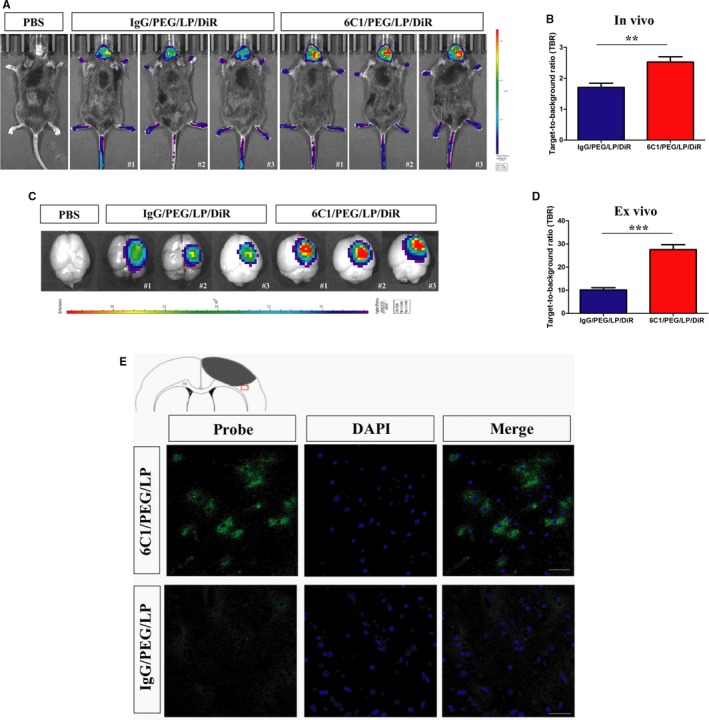
Representive in vivo NIRF images (A) and quantification of target‐to‐background (TBR) values (B) of ischemic mice after injection of immunoliposome nanoprobes at 24 hours. Brain tissues were dissected and acquired NIRF imaging at 24 hours after injection (C). Quantification of TBR values of brain tissues (D). Data are the mean±SEM (n=5 in PBS group or n=6 in nanoprobe groups). Statistical significance was indicated by: **P<0.01: ***P<0.001. E, Representive ex vivo fluorescence microscopy images of the peri‐infarct region of cerebral cortex ischemic mice obtained 24 hours after intravenous injection of anti‐PirB vectorized liposomes or control liposomes (cell nuclei stained in blue with DAPI; scale bar=50 μm). PirB, paired immunoglobulin‐like receptor B.
