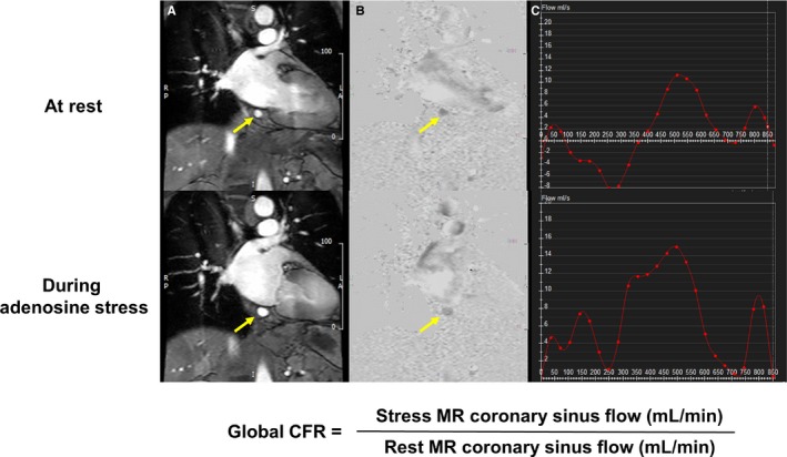
Figure 2.

Measurement of blood flow in the coronary sinus. Phase‐contrast cine magnitude image (A), velocity map (B), and blood flow curve in the coronary sinus (arrows) at 1 cardiac cycle (C). CFR indicates coronary flow reserve; MR, magnetic resonance.
