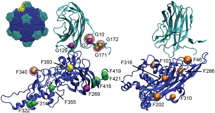
Fig. 2.
Locations of observed mutations in G4 lineages (left) and WA13 (right). Coat protein (F) shown in blue, spike protein (G) shown in teal. Full capsid is shown in the top left, with a single copy each of F and G highlighted in yellow. Of the G4 lineages, the sites of mutations that fixed in ID11 are shown in green, mutations in ID8 are shown in yellow, and mutations in NC13 are shown in purple. A purple halo around a yellow site indicates a site shared between ID8 and NC13. G4 mutations are significantly clustered, with the first-, second-, and third nearest neighbors of each mutation being significantly closer together than expected for a set of random mutations.

