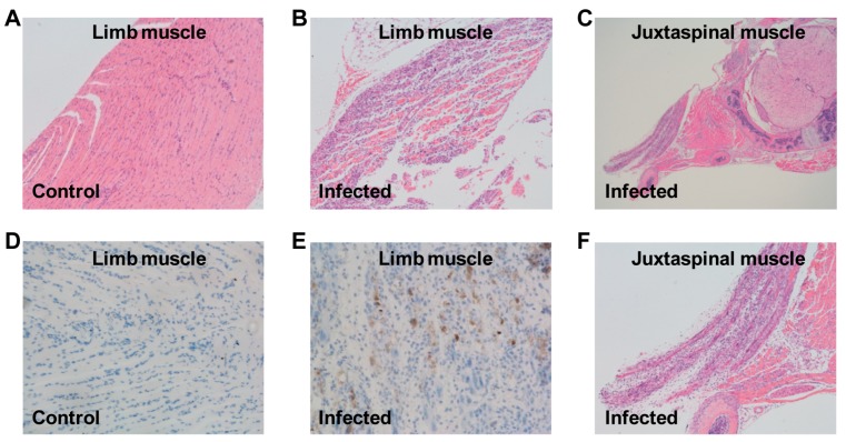
Figure 5.
Histological examination of EVD68-infected mice. One-day-old ICR mice were inoculated i.p. with (A,D) culture medium or (B,C,E,F) 1.2 × 10⁵ TCID50 of US/MO/14-18947. Tissues were collected at day 7 after inoculation and then subjected to (A–C,F) H&E staining or (D,E) immunohistochemical staining with an anti-EVD68 mAb 6A11 as the primary antibody; (F) expanded view of panel (C). Magnification: panels (A,B,F): ×100; panel (C): ×40; panels (D,E): ×200. Images shown are representative of two ICR mice in each group.

