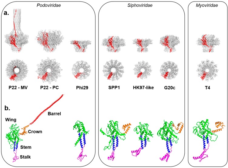
Figure 1.
Crystal structures of in vitro assembled portal proteins deposited in the RCSB Protein Data Bank. (a) Quaternary structures of portal proteins from Podoviridae, Siphoviridae and Myoviridae shown as side and top views. All structures are aligned with respect to the Mature Virion (MV)-portal protein of P22 and are shown in scale with the protomer “A” colored in red and the rest of the oligomer in gray; (b) Tertiary structures of portal protein protomers color-coded to highlight the stalk (magenta), stem (blue), wing (green), crown (orange) and barrel (red). Except for P22 portal protein, which is shown only in the MV-conformation, all other portal protomers are in scale.

