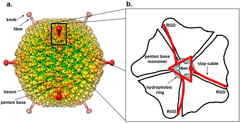
Figure 3.
Symmetry mismatch between trimeric fiber and pentameric penton base of adenovirus. (a) Surface rendered view of 3D-dimensional reconstruction of human adenovirus 26 (EMD-8471). (b) Schematic model for the attachment of adenovirus trimeric fiber to the penton base. The fiber is drawn as a red triangle with extended stay-cables interacting asymmetrically with the pentameric penton base (in black). The schematic diagram is drawn as proposed by Hong Zhou and collaborators in [178]. Hydrophobic residues in the penton base located at the rim of the channel are shown as a blue circle.

