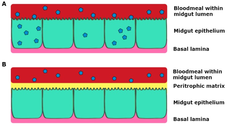
Figure 3.
Schematic representation of infection of the midgut, where virus particles are represented as blue polygons. (A) Virus in the bloodmeal infects epithelial cells through the microvilli and replicates; (B) Virus in the bloodmeal fails to infect the epithelial cells prior to secretion of the peritrophic matrix by midgut epithelial cells during blood digestion.

