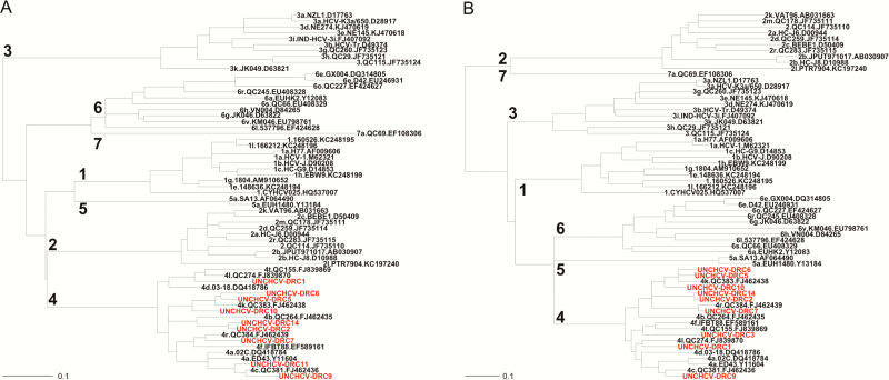
Figure 3.
Phylogenetic analyses. Samples from the current study (red) clustered with reference sequences (black) from known genotype 4 subtypes for both the hepatitis C virus NS5B (A) and core/E1 (B) regions, except for samples 2 and 14. Reference sequence identifying information is separated by periods and includes the following (from left to right): genotype and subtype, isolate identifier, GenBank accession number.

