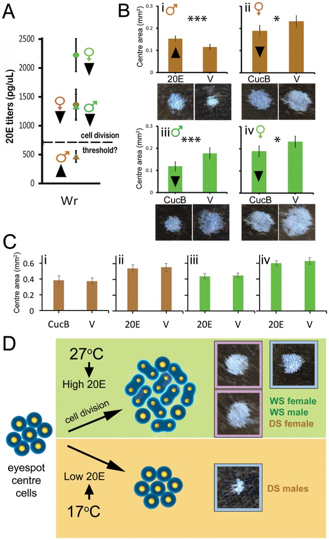
Fig. 4.

20E signaling promotes an increase in eyespot center size. (A) 20E titers in developing larvae at end of Wr stage. Dashed line represents hypothetical threshold of 20E titers required for cell division. Arrowheads next to data points represent planned manipulations to 20E signaling. (B) 20E injections cause an increase in eyespot size in DS males (i), whereas reduced EcR signaling using CucB causes a decrease in eyespot size in all other groups (ii–iv). Figures below respective graphs represent representative images obtained after treatments. (C) Opposite-direction hormone treatments (to the arrowheads in (A)) does not produce any significant differences in DS males (i), DS females (ii), WS males (iii), and WS females (iv), supporting the threshold-response hypothesis for cell division. Error bars represent 95% CI of means. (D) Diagram summarizing the interpretation of our results: Rearing temperature induces variation in 20E titers at the Wr stage of development. High titers result in cell division and larger eyespot centers, whereas low titers result in smaller centers, as seen in DS males (blue outlines = males; pink = females). DS females, despite being reared at low temperature, have sufficiently high 20E levels to also undergo cell division of the wing ornament.
