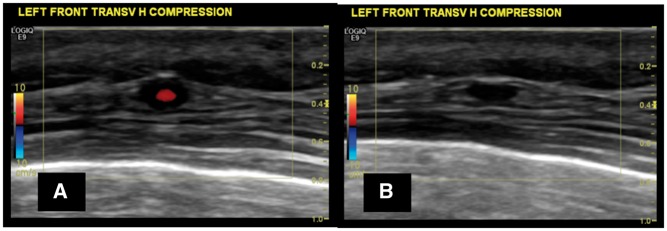
Fig. 1.
US image showing a halo around the temporal artery and a positive compression sign
(A) Halo sign at the level of the frontal branch of the right temporal artery, transverse view, before applying compression, in a patient with active GCA. (B) Evidence of a positive compression sign with the halo persisting despite firm compression applied with the transducer.

