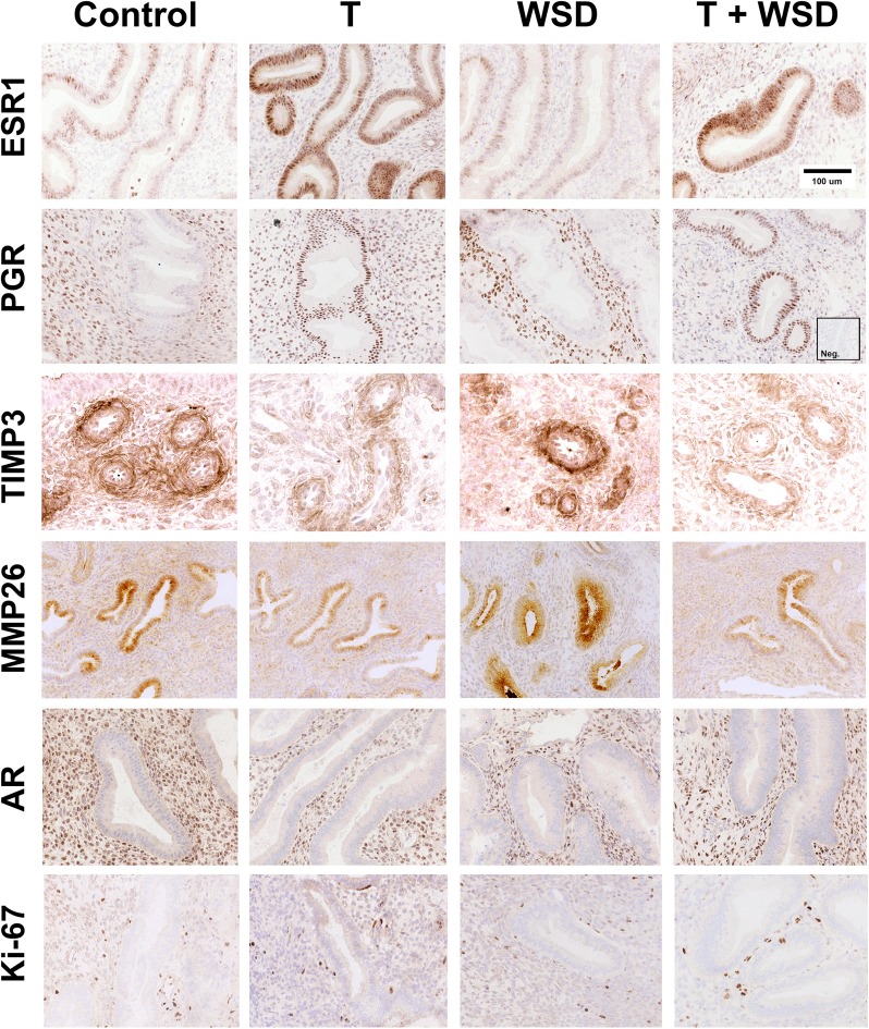
Figure 5.
Photomicrographs illustrating immunohistochemical staining for estrogen receptor 1 (ESR1), progesterone receptor (PGR) MMP26, TIMP3, Ki-67 and androgen receptor (AR) in the endometrial functionalis zone of the macaque uterus from representative females in each treatment group (C, T, WSD, T+WSD). Brown staining denotes positive expression of proteins. Sections are counterstained with hematoxylin (blue) staining. ESR1, PGR, Ki-67 and AR staining is nuclear, while MMP26 and TIMP3 show cytoplasmic localization. Inset shows a negative control with an irrelevant antibody (Anti-Br(d)U). TIMP3 staining was localized to the predecidual cells around the spiral arteries.

