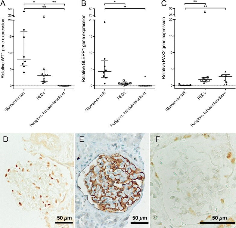
Fig. 3.

Expression levels of selected transcripts in micro-dissected non-injured hemalaun-stained glomerular substructures. Nine cases with acute tubular injury and mild interstitial inflammation, but without any glomerular disease were used (cohort I). WT1 (A), GLEPP1 (B) and PAX2 (C) seem to be reliable discriminators for proper micro-dissection of glomerular tufts vs. PECs. For comparison, periglomerular tubulointerstitium is shown. There were significant differences between glomerular tufts and PECs regarding all markers. Relative expression was calculated using the geometric mean of PGK1 and GAPDH as normalization factor. There was no expression of WT1 and GLEPP1 (beside one outlier) in periglomerular tubulointerstitium. In one patient case, no amplification of target and reference transcripts was detectable in PECs. One outlier was excluded in WT1 and GLEPP1 transcript expression in glomerular tufts. p-values were calculated by the Wilcoxon matched-pairs signed rank test (*p < 0.05; **p < 0.01). For comparison, immunostaining of WT1 (D), GLEPP1 (E) and PAX2 (F) is given in one selected case. WT1 (D) and GLEPP1 (E) are strongly expressed in podocytes and to a lower extend also in PECs, but not in the tubulointerstitium. PAX2 is expressed in PECs and tubular epithelial cells (F). Magnification ×400 or ×200
