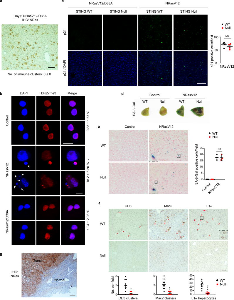
Extended Data Figure 6. STING promotes Ras-induced SASP in the liver.
a, Immunohistochemistry of WT liver injected with NRasV12/D38A mutant. b, Hepatocytes of injected WT mice were isolated on day 6 and stained. CCF-positive hepatocytes were quantified. Results are average values of four different fields with over 200 cells; *p<0.001, compared to control and NRasV12/D38A. c, Liver was analyzed on day 6 for p21. n=4 mice. d–e, SA-β-Gal analyses of liver on day 6. n=3 mice, mean with s.e.m for e. f, Liver was analyzed by immunohistochemistry on day 6 and quantified. n=8 mice; *p<0.005, **p<0.001, ***p<0.0005. g, Liver tumor stained for NRas. One-way ANOVA coupled with Tukey’s post hoc test (b) and unpaired two-tailed Student’s t-test for all others. Scale bars: 10 µm for b and 100 µm for all others. Error bars are s.e.m.

