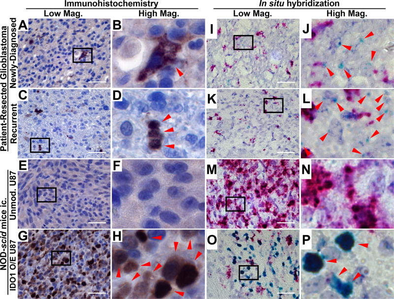
Figure 1. Immunohistochemical (IHC) IDO1 localization and in situ hybridization (ISH) for IDO1 in human glioblastoma (GBM).
(A–H) IHC detection with a human-specific IDO1 antibody was performed in (A, B) newly diagnosed primary patient-resected GBM, (C, D) recurrent patient-resected GBM or tumors isolated from immunodeficient NOD-scid mice intracranially-engrafted (ic.) (E, F) unmodified U87 cells and (G, H) U87 overexpressing human IDO1 conjugated to an HA tag (HA-IDO1 O/E). A positive signal is indicated by red arrows in the high magnification images (B, D, F, H). (I–P) ISH for human IDO1 mRNA was performed in the same tissues described for IHC and is indicated with red arrows in the high magnification images (J, L, N, P). The proliferation marker, Ki67, was used as a positive control and appears as a red signal. All lower magnification images were obtained under 64× objective lens with a scale bar representing 400 µm.

